Center for Translational Imaging to Capitalize on Georgetown’s History and Expertise in Imaging Science
(December 7, 2018) — The ability to peer inside the body to learn if something has gone wrong and if so, what, is one of the critical pillars of health care today, but as a team of researchers have long realized, the field of imaging provides a powerful way to reveal unimagined clues to better understand health and disease in the laboratory.
Driven by that conviction of the power of imaging, Christopher Albanese, PhD, led a successful effort to establish the Georgetown Center for Translational Imaging (CTI), announced this fall.
The CTI combines the revealing forces of the Preclinical Imaging Research Laboratory (PIRL), established by Albanese, and the Center for Functional and Molecular Imaging (CFMI), establish by John VanMeter, PhD, who will also help lead the CTI.
The CFMI is oriented to human and, to a lesser degree, some preclinical research, while the PIRL is the backbone of small animal imaging and biophysical research projects.
“As the only center of its kind in the Washington, D.C., area, the Center for Translational Imaging will integrate complementary clinical and preclinical imaging research approaches to provide a unique opportunity to solidify Georgetown’s stature in MR imaging,” says Albanese, professor of oncology and pathology.
Historical Roots
“The CTI will also integrate the combined knowledge gained in the past 25-plus years in the field of imaging hardware and software development,” adds Albanese. “That knowledge can be applied to the fields of biomedical engineering, developmental biology, drug discovery, oncology, neurology, neuroscience and infectious disease, among others.”
The new center builds on a legacy established decades ago with the development of the first CT scanner.
Developed at Georgetown in the mid-1970s by the late Robert S. Ledley, DDS, the technology was first used to evaluate the injuries of a 4-year-old boy who reportedly had fallen off his bike and hit his head. The scanner visualized a blood clot in the child’s brain that could have been fatal had it not been for the surgery that quickly followed the scan.
The CFMI
Today, neuroimaging at Georgetown holds a strong place in non-clinical research. The CFMI and its staff of experts are leaders in the application of advanced neuroimaging techniques to better understand such disorders as Alzheimer’s disease, stroke and epilepsy. Other studies use neuroimaging to research chronic fatigue syndrome, HIV-associated neurocognitive disorders, Gulf War illness and the effect on the brain of the chemotherapy used to treat breast cancer. A significant percentage of the research conducted at CFMI focuses on adolescent development, such as addiction research and disorders including autism and attention deficit hyperactivity disorder.
The CFMI, created in 2003, currently supports 37 active principal investigators from eight different institutions. Of their projects, 25 are supported by NIH grant funding totaling more than $11 million.
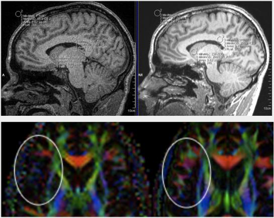
Efforts to increase Georgetown’s imaging capacity have led to equipment upgrades. This past fall, the CFMI upgraded its 3 Tesla Siemens Magnetom Tim Trio MRI scanner to provide crystal-clear neuroimaging. The NIH funded the “PRISMA” upgrade with more than $1.3 million in support.
“The quality of images is much higher,” says VanMeter. “We have more detail, higher spatial resolution and the scan can be done more quickly. It’s fantastic.”
The PIRL
Founded by Albanese in 2006, the PIRL supports 18 projects primarily funded by the NIH, in addition to studies supported by the Department of Defense and the National Science Foundation. Grant funding exceeds $11 million, with projects covering a wide range of topics including oncology, neurobiology, infectious diseases and contrast agent development.
The PIRL provides the research community with access to state-of-the-art non-invasive in vivo imaging technologies for the characterization of animal models of disease and for studies that involve the chemical and physical evaluation of experimental phantoms and contrast agents.
“Our work in the PIRL provides a strong basis for translational work,” Albanese says. Moreover, the PIRL offers a comprehensive program that facilitates image acquisition and advanced and quantitative image analysis to advance research in fields such as childhood and adult cancers, spinal cord injury and repair, and bacterial infections.
Collaboration Expansion
The CTI will collaborate with the National Institutes of Health (NIH), Lawrence Livermore National Laboratory and other institutions, including Columbia University, Mass General Hospital in Boston and the University of Maryland, says Albanese. It will also serve other local, national and international academic and medical research communities and interact with government agencies, pharmaceutical companies and private foundations.
Additionally, the CTI is planning to develop graduate level education at Georgetown in medical physics, says Albanese, something that the D.C. region currently lacks. To that end, Georgetown has recruited Stanley Fricke, PhD, who will return to the Hilltop in 2019 to serve as the CTI’s director of imaging and education (his most recent position was at Children’s National Medical Center).
“Dr. Fricke played an integral role in the PIRL and CFMI, and we’re pleased he’ll return to lead medical physics and engineering graduate education,” says Albanese.
“We see the CTI as a hive of imaging benchwork, education and engineering,” Albanese adds. “It will secure Georgetown’s spot on the medical and scientific imaging map — a place where we belong.”
GUMC Communications