A Brain Fingerprint: Study Uncovers Unique Brain Plasticity in People Born Blind

Posted in News Release | Tagged blindness, brain, brain development, brain plasticity, neuroscience
Media Contact
Karen Teber
km463@georgetown.edu
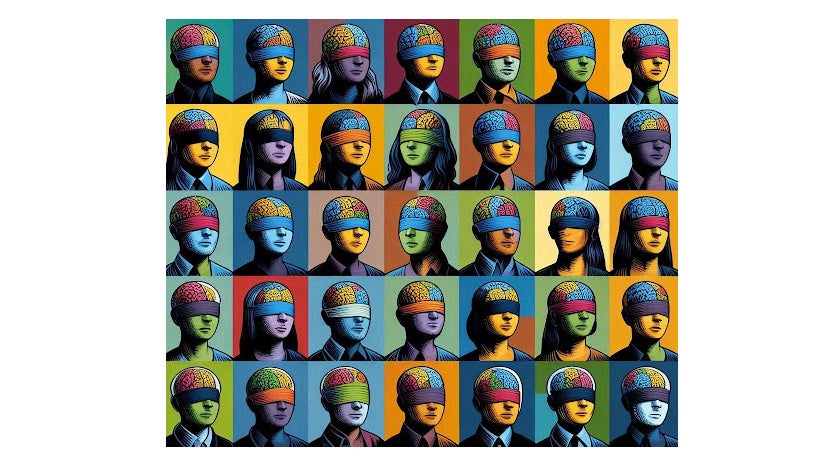
WASHINGTON (July 30, 2024) — A study led by Georgetown University neuroscientists reveals that the part of the brain that receives and processes visual information in sighted people develops a unique connectivity pattern in people born blind. They say this pattern in the primary visual cortex is unique to each person — akin to a fingerprint.
The findings, described July 30, 2024, in PNAS, have profound implications for understanding brain development and could help launch personalized rehabilitation and sight restoration strategies.

For decades, scientists have known that the visual cortex in people born blind responds to a myriad of stimuli, including touch, smell, sound localization, memory recall and response to language. However, the lack of a common thread linking the tasks that activate primary areas in the visual cortex has perplexed researchers. The new study, led by Lenia Amaral, PhD, a postdoctoral researcher; and Ella Striem-Amit, PhD, the Edwin H. Richard and Elisabeth Richard von Matsch Assistant Professor of Neuroscience at Georgetown University’s School of Medicine, offers a compelling explanation: differences in how each individual’s brain organizes itself.
“We don’t see this level of variation in the visual cortex connectivity among individuals who can see — the connectivity of the visual cortex is usually fairly consistent,” said Striem-Amit, who leads the Sensory and Motor Plasticity Lab at Georgetown. “The connectivity pattern in people born blind is more different across people, like an individual fingerprint, and is stable over time — so much so that the individual person can be identified from the connectivity pattern.”

The study included a small sample of people born blind who underwent repeated functional MRI scans over two years. The researchers used a neuroimaging technique to analyze neural connectivity across the brain.
“The visual cortex in people born blind showed remarkable stability in its connectivity patterns over time,” Amaral explained. “Our study found that these patterns did not change significantly based on the task at hand — whether participants were localizing sounds, identifying shapes, or simply resting. Instead, the connectivity patterns were unique to each individual and remained stable over the two-year study period.”
Striem-Amit said these findings tell us how the brain develops. “Our findings suggest that experiences after birth shape the diverse ways our brains can develop, especially if growing up without sight. Brain plasticity in these cases frees the brain to develop, possibly even for different possible uses for the visual cortex among different people born blind,” Striem-Amit said.
The researchers posit that understanding each person’s individual connectivity may be important to better tailor solutions for rehabilitation and sight restoration to individuals with blindness, each based on their own individual brain connectivity pattern.
The authors report having no personal financial interests related to the study.
This work was supported by a grant from National Institutes of Health NEI/OBSSR (R01EY034515) and funds from the Edwin H. Richard and Elisabeth Richard von Matsch Distinguished Professorship in Neurological Diseases.
