Keeping Cells Alive in the Lab
Posted in GUMC Stories | Tagged cancer, cancer research
(January 19, 2012) — There is a lot of talk about personalized cancer treatment in medicine these days. One idea is to subject a tumor to different therapies to find the cocktail of drugs that will work with limited toxicity — well before a patient actually is treated. It’s what many oncologists look forward to — someday.
But it recently happened in a lab at Georgetown Lombardi Comprehensive Cancer Center. A donated sliver of a patient’s lung tumor was grown into numerous small tumors in a laboratory and then they were systemically exposed to various cancer drugs to see which agents offered the most potent killing potential. The best one was then used to treat the patient.

This advance was possible because a team of 13 Georgetown researchers, with a hand from several scientists at the National Cancer Institute, have figured out how to keep both normal and cancer cells alive in the laboratory — which previously had not been possible.
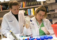
Normal cells usually die in the lab after dividing only a few times, and many common cancers will not grow, unaltered, outside of the body, says Richard Schlegel, M.D., Ph.D., chairman of the department of pathology at Georgetown Lombardi.
But now the new technique, described December 19 in the American Journal of Pathology, can keep these cells alive indefinitely. This breakthrough could not only usher in a new era of personalized cancer medicine, it has potential application in regenerative medicine, as well as in decoding the molecular differences between a patient’s normal and tumor cells, says Schlegel.
“We discovered we can grow normal and tumor cells from the same patient forever, and nobody has been able to do that,” he says. “Normal cell cultures for most organ systems can’t be established in the lab, so it wasn’t possible previously to compare normal and tumor cells directly.

“We tried breast cells and they grew well. We tried prostate cells and their growth was fantastic, which is amazing because it is normally impossible to grow these cells in the lab,” Schlegel says. “We found the same thing with lung and colon cells that have always been difficult to grow.”
The ability to grow true cancer cells may revolutionize basic science as well, Schlegel says, because many of the cancer cell lines used for basic research in labs worldwide have accumulated genetic changes that don’t resemble the original primary tumors.
The research team found that adding two different substances to cancer and normal cells in a laboratory pushes them to morph into stem-like cells — adult cells from which other cells are made.
The two substances are a Rho kinase (ROCK) inhibitor and fibroblast feeder cells. ROCK inhibitors help stop cell movement, but it is unclear why this agent turns on stem cell attributes, Schlegel says. His co-investigator Alison McBride, Ph.D., of the National Institute of Allergy and Infectious Diseases, had discovered that a ROCK inhibitor allowed skin cells (keratinocytes) to reproduce in the laboratory while feeder cells kept them alive.
The ability to immortalize cancer cells will also make biobanking — storing tumors for both research and to improve individual treatment — both viable and relevant, Schlegel says.
The researchers further discovered that the stem-like behavior in these cells is reversible. Withdrawing the ROCK inhibitor forces the cells to differentiate into the adult cells that they were initially. This “conditional immortalization” could help advance the field of regenerative medicine, Schlegel says.
However, the most immediate change in medical practice from these findings is the potential they have in “revolutionizing what pathology departments do,” Schlegel says.
“Today, pathologists don’t work with living tissue. They make a diagnosis from biopsies that are either frozen or fixed and embedded in wax,” he says. “In the future, pathologists will be able to establish live cultures of normal and cancerous cells from patients, and use this to diagnose tumors and screen treatments. That has fantastic potential.”
The technique works now with epithelial cancers — breast, prostate, colon, and liver — but it could potentially be used for any disease that affects these kinds of tissues. “You could compare a patient’s liver disease to normal liver cells to figure out what is going awry, or examine the effect of cystic fibrosis on lung tissue, compared to normal lung,” Schlegel says. “The uses are endless.”
Word of the achievement spread even before the study was published, and Schlegel has already shown representatives from several universities who came visiting just how it works. He says investigators can use the method for research purposes.
“If this advance helps them understand the roots of disease, and improve treatment, it helps us all,” Schlegel says.
By Renee Twombly, GUMC Communications
