Art Installation Illustrates Dyslexic Brains at Work
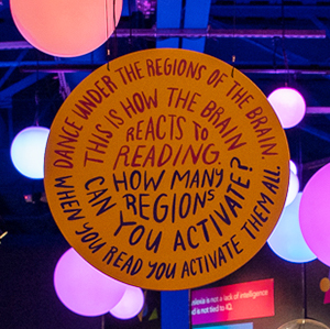
(January 14, 2021) — When a person practices a skill, the neural representations in the relevant parts of the brain change, allowing the person to perform the skill better. Research by Guinevere Eden, PhD, a Georgetown University professor of pediatrics and the director of the Center for the Study of Learning, found that the same kind of brain plasticity occurs when people receive coaching and practice in reading.
“We’ve learned that there is a specialization of areas in the visual system for words in adults which has yet to develop in children,” Eden says. “You can literally see how the educational experience changes our brains to be better attuned to words.” Eden is a physiologist who uses functional magnetic resonance imaging, or fMRI, to see which areas of the brain are being used in children and adults when they read; and also how they differ in those children and adults who have the reading disability dyslexia.
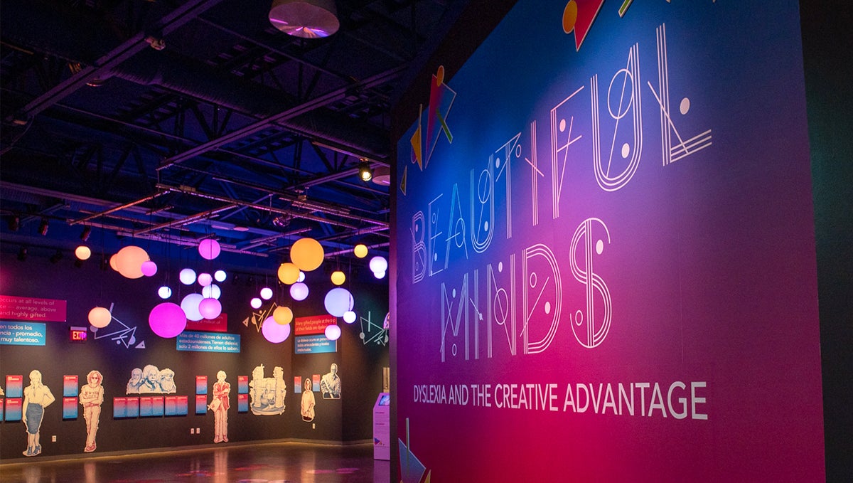
An interactive light sculpture installed earlier this year at a children’s museum draws on Eden’s research and makes the brain’s representation of reading visible. Sarah Sudhoff, a photojournalist and artist, was selected in August to be artist-in-residence at the DoSeum in San Antonio, Texas. When the museum asked her to create an installation in the center of an exhibit about dyslexia, Sudhoff was inspired by her 8-year-old son, who has dyslexia, to create something that would illustrate how dyslexic brains work.
Science Inspires Art
Sudhoff had read about Eden’s work while researching dyslexia, and decided to ask Eden for data to use as the basis of the installation. Though Eden had never had connected her research to art, “I loved the idea,” Eden says. “I grew up in an art-loving home and as a child met many artists, including Henry Moore and Salvador Dali.”
A photojournalist and former photo editor for Time magazine, Sudhoff says her art projects “are still based in reality and research, revealing something to the public in a truthful way.” Her earlier works have incorporated patient heart rates and respiration during air ambulance trips and images of cells infected with bubonic plague.
Eden and postdoctoral fellow Anna Matejko, PhD, recently conducted a study in children with dyslexia that used fMRI to measure brain activity during two four-minute scans while the children viewed real and made-up words. They sent the activation measures from some of their brain regions for a subset of unidentified children to Sudhoff.
From Brain Scans to Interactive Artwork
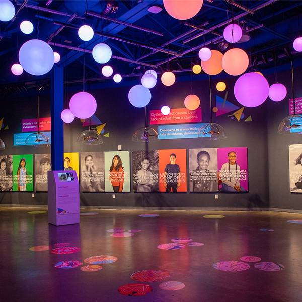
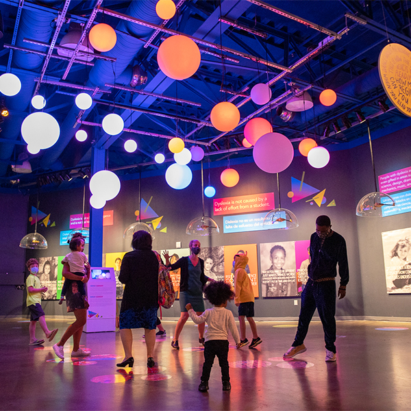
With the help of a computer programmer, Sudhoff used the data to make 36 acrylic orbs, representing abstract brain areas that light up in an intensity that correlates with the children’s brain signals while processing words. The orbs are suspended from the ceiling in the center of the exhibit space, loosely arranged into the brain’s two hemispheres, and symbolize brain regions known to be involved in reading. As people move beneath them, additional changes occur: The intensity of color and frequency of flashes increase in response to what the museum’s visitors are doing below.
“When you have three people or more, the colors get more saturated and move more quickly,” Sudhoff says. The more activity the visitors generate, whether by running, dancing or flailing their arms, the stronger the color of orbs becomes. The deeper red and orange “visually signify the more you read, the stronger your neural activity becomes,” Sudhoff says.
Searching For the Changes That Lead to Reading Improvement
Eden and her colleagues are currently analyzing the data from their study, which involved a series of brain scans acquired before and after the children with dyslexia received tutoring that improved their reading ability. “We are interested in understanding the brain’s response to a training experience that drove their reading gains,” Eden said. That is, which specific brain regions are increasing or decreasing their activity following the intervention.
“When children with dyslexia read, those brain regions that are activated in children who read well are found not to be activated as much in children who have dyslexia,” says Eden. “One thing that we have seen in a prior study in adults with dyslexia, is that in addition to seeing increases in brain activity in these regions following a reading intervention, there are brain areas outside of these regions that become more active, indicative of compensation. It’s similar to what is reported in stroke patients, where a region takes over the job of its neighbor,” Eden says.
A retraining process actually happens in everyone’s brain when they learn to read, Eden says. Humans have only been reading for about 5,000 years, and as such there are no brain regions dedicated to this skill from an evolutionary perspective. The areas of the brain that we use for reading were originally designed to do something else and then got recycled for reading, she says.
The “Reading Brain” installation will remain on display in the Beautiful Minds exhibit at the DoSeum through March 28.
Kathleen O’Neil
GUMC Communications