
Uncovering Clues in the Brain
Posted in GUMC Stories
 It’s been said the human brain is the ultimate supercomputer – the wiring is so complex that no machine can rival it. It’s true – the top 1/5 of an inch of the brain surface contains billions of neurons, each linked to thousands of other neurons through their long axons. These axons provide highways that snake through the brain sideways, upwards, from front to back, and back to front.
But unlike a computer, whose circuits are all interchangeable, the brain’s processing centers (the gray matter) and the information highways (white matter) can be quite variable. No one’s brain is exactly alike, but large differences caused by genetics, disease, or trauma can contribute to neurological impairment.
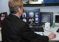
So say the array of researchers imaging human brains at the Center for Functional and Molecular Imaging at Georgetown University Medical Center. These scientists and their participants form a steady current in and out of the center, located in the Preclinical Science Building.
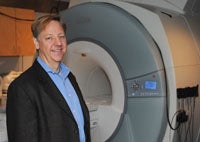
There lies a white and gray whole-body functional MRI (fMRI) scanner, the most powerful available, that is complete with an embedded TV and DVD, that is overseen by a research staff of nine, which includes the center’s director John VanMeter, Ph.D.
Observing the Brain in Action
Researchers can watch, in real time, what happens inside the brain given certain stimuli. fMRI measures change in blood flow and blood volume due to cognitive activity in the brain. The imaging isn’t invasive and doesn’t emit radiation. The only trick is that participants have to lie still while the scanner captures changes in the magnetic fields of protons.
“The brain is a beautiful thing, especially when you can see it in action,” says VanMeter.
At the center, researchers are figuring out the neural basis of everything from language development to face recognition, and how these skills may impact disorders such as autism and dyslexia. They are also trying to piece together the neural back-story to disorders that range from fibromyalgia and Gulf War illness to epilepsy.
There are also studies that use brain imaging to explore such behavior as empathy and deception and others that probe thought and feeling.
A Research Resource for Metropolitan Washington
Since 2005, nearly 5,000 MRI sessions have been held at the center with 2,172 study and control participants, both adults and children, some of whom are as young as four years old.
Outside of the National Institutes of Health (NIH), the center offers the only research-dedicated fMRI machine in the greater Washington, D.C. area. It supports researchers from eight departments at Georgetown, from two campuses, as well as those from George Washington University, George Mason University, Children’s National Medical Center, and American University.
The number of studies for each week may vary but probably averages around 15, VanMeter says. Georgetown funded the scanner, the facility renovation, and computation infrastructure for about $3 million, and the center received support from NIH.
The use of fMRI in brain research is relatively new – the first successful study using the technology was reported in 1991, and VanMeter, having just obtained his degree in computer science from Dartmouth University, happened to be a staff fellow at the NIH doing a different kind of imaging when fMRI started taking off in a big way.
Now the Georgetown facility forms the basis upon which a wide variety of studies are designed, all with an ultimate goal of developing improved tools for diagnosis and treatment.
A Window into Autism
Autism is a subject several researchers study, including VanMeter. He has found differences between autistic individuals and those without the disorder in both the structure of the brain and in activity in the amygdala, the home of emotion. For example, VanMeter has found that parts of the corpus callosum, the wide bundle of axons that connects the left and right cerebral hemispheres, are smaller in some children with autism. And his research suggests that narrowing occurs in different areas according to the subtype of autism. Additionally, the amygdala seems to be hyperactive more often in autistic kids, he says.
Associate psychology professor Chandan Vaidya, Ph.D., has also discovered differences in information flow. “We have found that how frontal lobe regions communicate with other regions differs based on the age of the autistic child – communication is reduced in older compared to younger children. In a study of typically developing children, we have found that communication across regions depends upon the integrity of the white matter connections between the regions.”
Researchers at both Georgetown and Children’s National Medical Center have found that deficits in face recognition likely contribute to the difficulties in social interaction that are one of the defining features of autism.
In some study participants with autism, neurons in an area of the brain known to be key to face recognition do not respond as selectively as in people without autism, says Maximilian Riesenhuber, Ph.D., a Georgetown neuroscientist and director of the Lab for Computation Cognitive Neuroscience. Other autistic participants, in contrast, may be using different areas of the brain for the task, resulting in a decreased ability to tell faces apart, he says.
“It may be that while the spectrum of autistic disorders often involve decreased ability to recognize faces, there are a number of ways this deficit can occur, and that varies across individuals,” says Riesenhuber. He adds that this finding may ultimately be helpful in providing more precise ways to characterize autism as well as leading to new strategies for personalized treatment.
At this point, the researchers cannot say how these brain regions came to be different, or even that these differences cause the disorder. “We don’t know what the origins of autism are, but my general take on it is that for unknown reasons, there are kids who are susceptible,” says VanMeter. “It could be an inherent susceptibly or an external trigger that causes a profound change.
“We also don’t have any idea what that trigger might be, but my bet is that it doesn’t happen after birth because that would essentially mean a thinking brain somehow rearranged itself,” VanMeter says, adding “There is much we don’t know about autism, but a few things that we do… thanks to our ability to take a little peek inside the most fabulous organ of all.”
By Renee Twombly, GUMC Communications
(Published January 19, 2012)